Ultrasound Diagnostic Systems Sonus

Portable ultrasound scanners for daily clinical practice: touchscreen controls and a wide range of imaging modes

Shipping within Ukraine

24-month official warranty on the system

Service, Parts Inventory, and Training in Ukraine

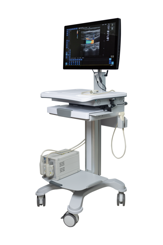
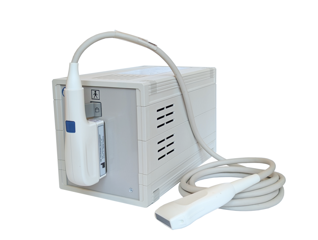
SONUS is a line of portable ultrasound scanning systems designed for a wide range of clinical applications: from abdominal imaging and obstetrics and gynecology to cardiology, vascular diagnostics, and musculoskeletal imaging.

The wideband beamforming system, color and energy Doppler, and elastography options provide clinical-grade image quality sufficient for most functional diagnostic tasks.

The compact, all-in-one unit with touchscreen controls significantly improves the convenience and speed of examinations.

Do you have any questions?

Get expert advice from our specialists

5 Advantages of SONUS Ultrasound Systems:

01 Many years of engineering experience

SONUS developers have been creating ultrasound diagnostic equipment in Ukraine for decades—a design tradition proven by time and the number of systems installed

02 Ukrainian manufacturer

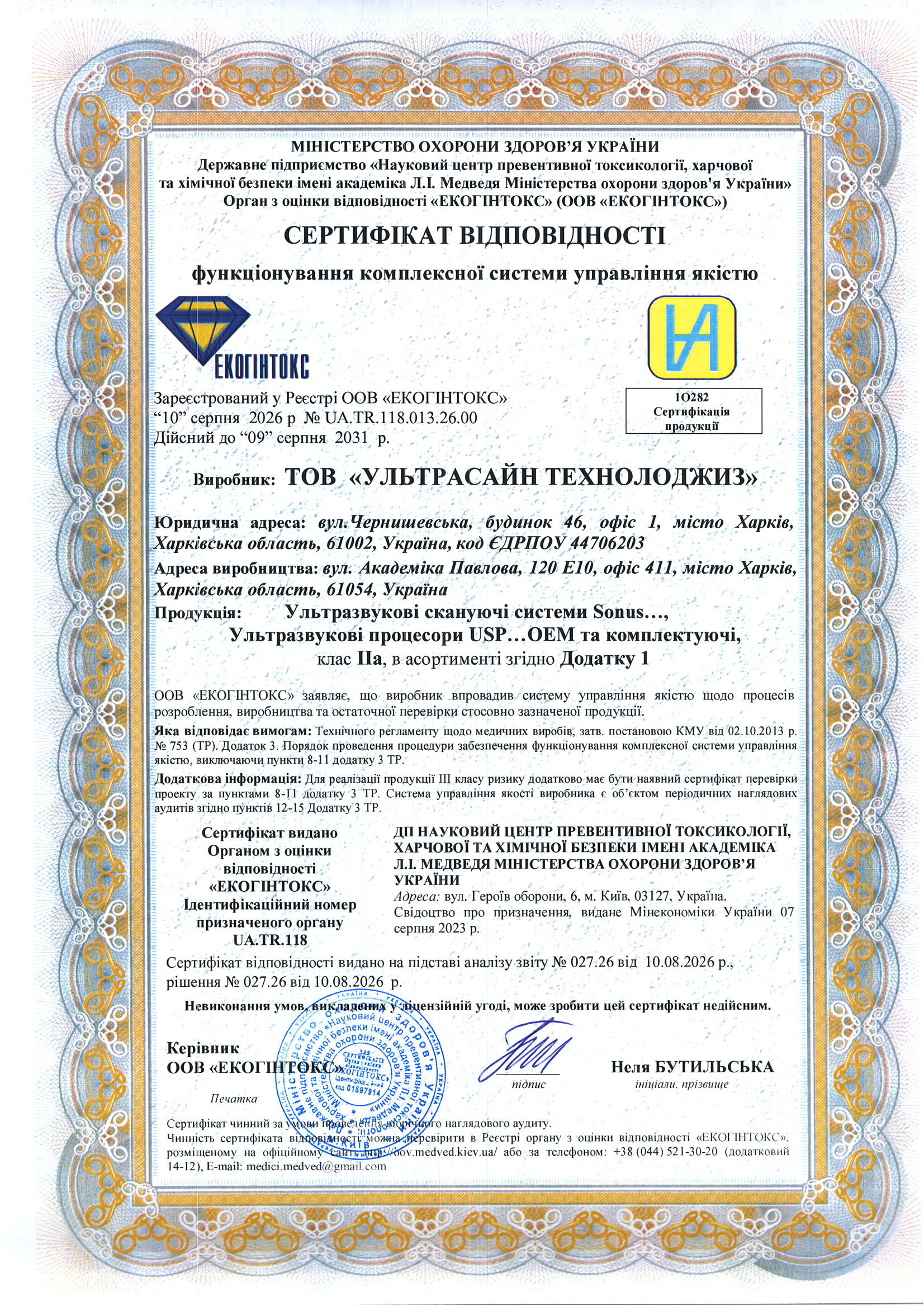
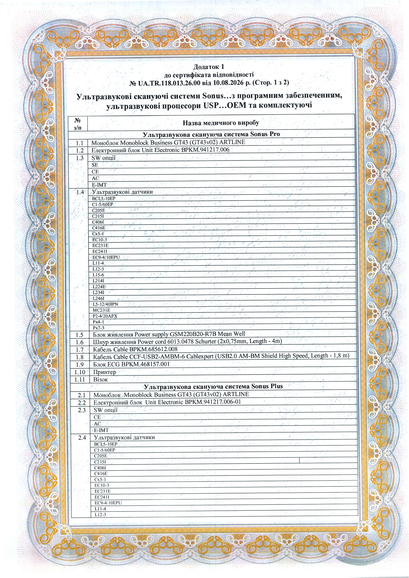
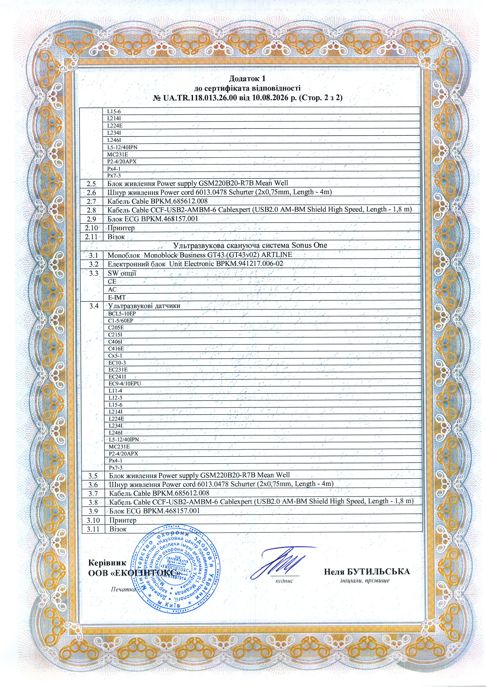
The equipment is manufactured in Ukraine and certified in accordance with the Technical Regulations for Medical Devices

03 Optimal value for money

Clinical functionality sufficient for the vast majority of daily examinations

04 Portability

This compact device, weighing no more than 2.5 kg (4.5 kg for pro models), is easy to transport for on-site examinations, field and veterinary practice, and can also be used in offices without specialized equipment

05 Touchscreen control

An intuitive touchscreen starting at 23.8″ reduces staff training time and minimizes the risk of errors when configuring examination modes

Clinical Applications of SONUS

One system—for most areas of ultrasound diagnostics in both general and highly specialized clinics:

🟠  Abdominal Organs
🟠  Gynecology and Obstetrics
🟠  Urology

🟠  Surgery

🟠  Vascular Imaging

🟠  Cardiology

🟠  Pediatrics and Neonatology

🟠 Anesthesiology and Intensive Care
🟠  Breast Medicine

🟠  Transcranial Imaging

🟠  Endocrinology

🟠  Oncology

🟠  Neurology

🟠  Musculoskeletal System

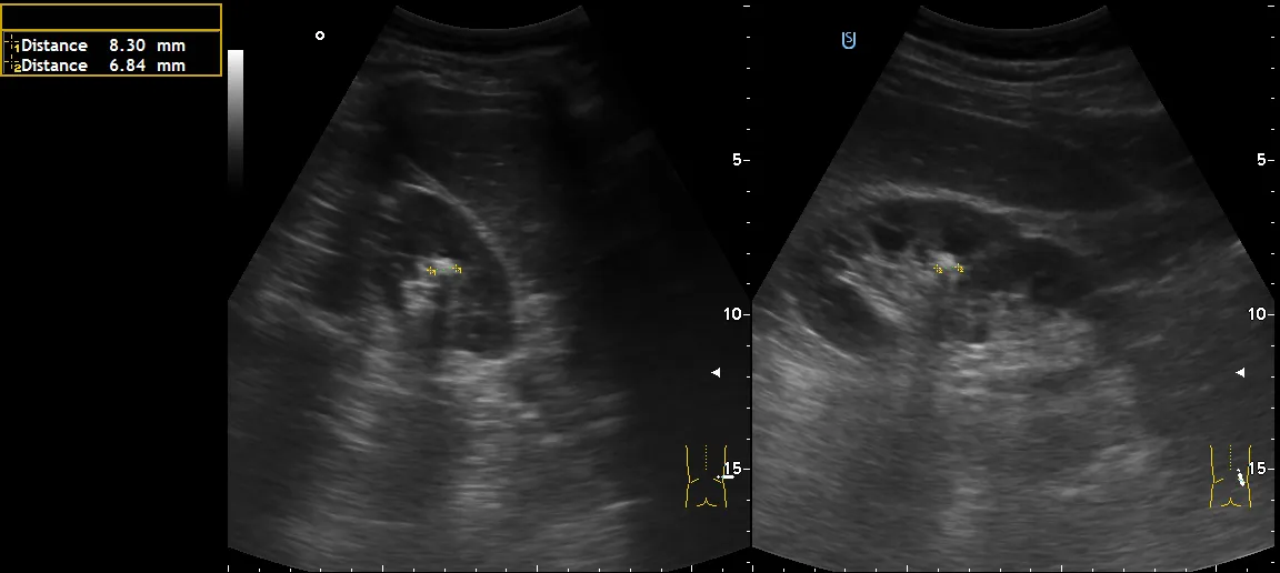
Right kidney
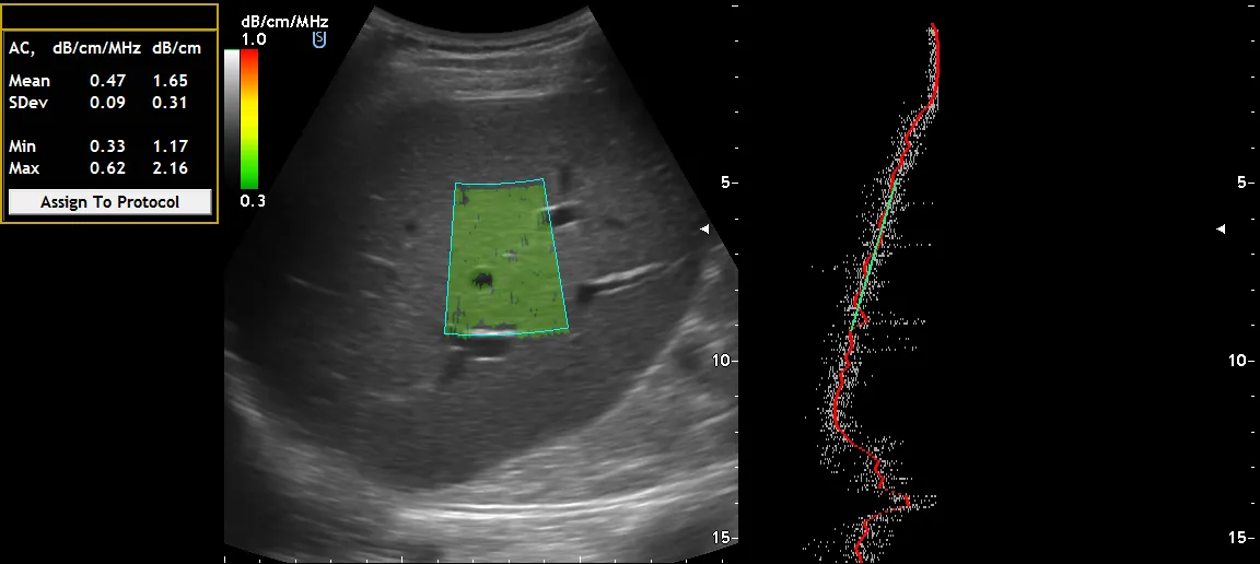
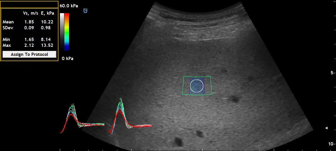
Liver
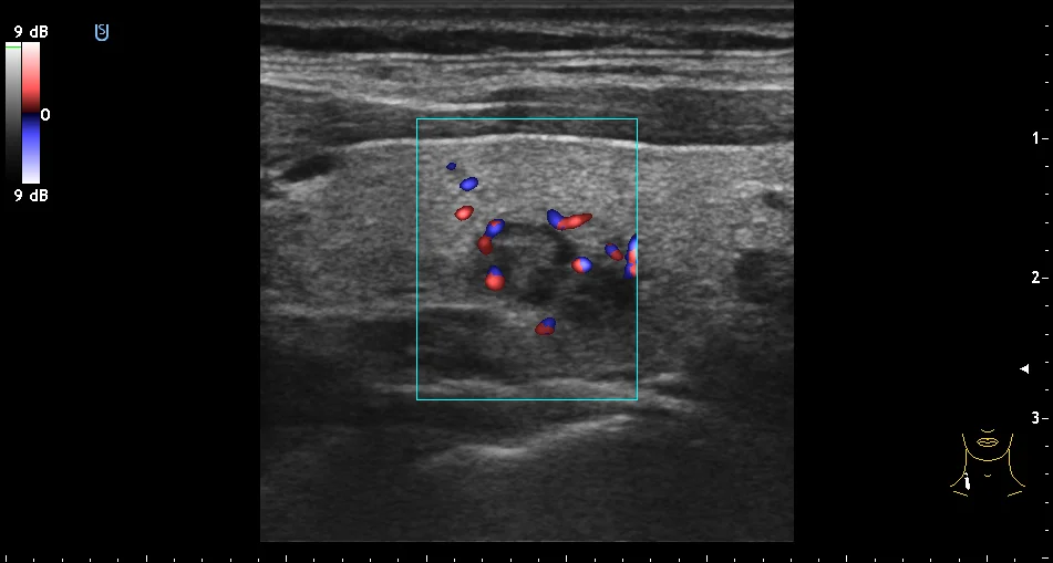
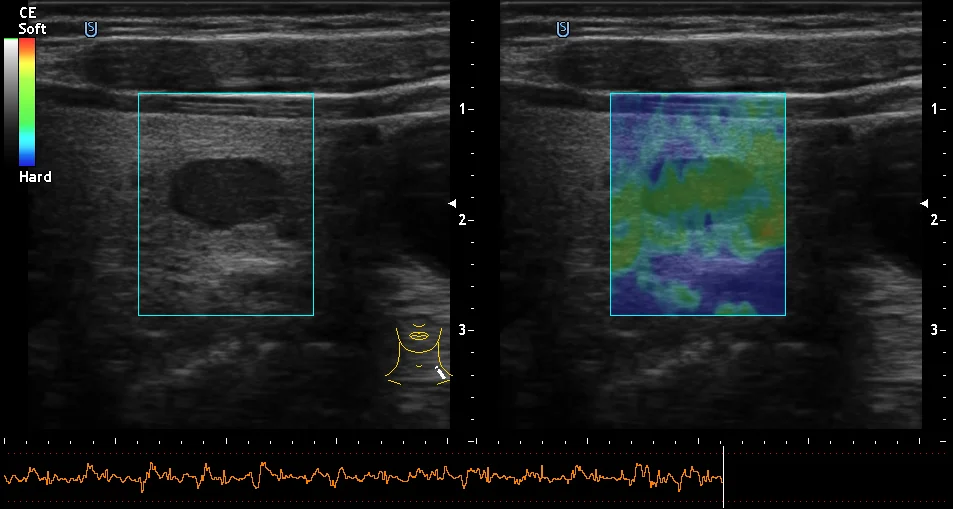
Thyroid Gland
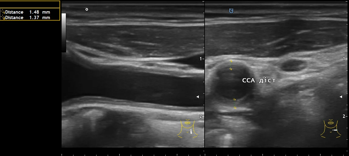
Carotid Artery
Thyroid Gland
Liver
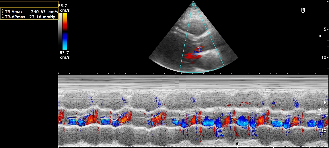
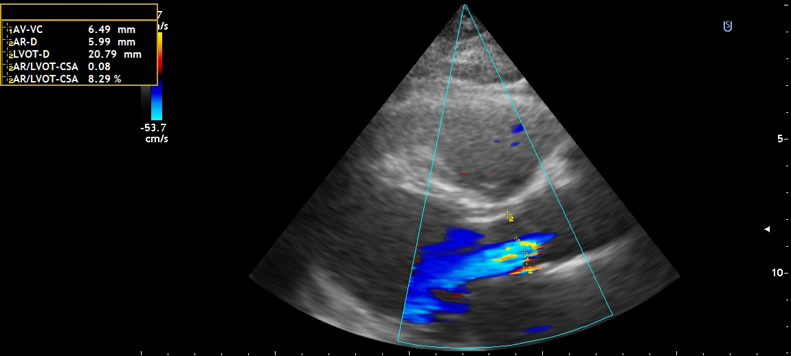
Aortic regurgitation
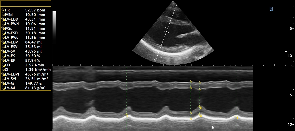
Heart
Aortic regurgitation in congenital heart disease
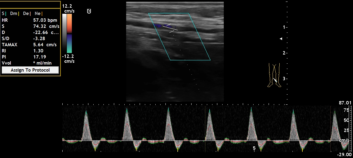
Blood Flow in the Tibial Artery
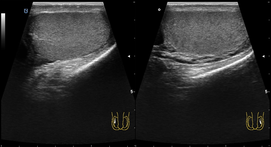
Testicles
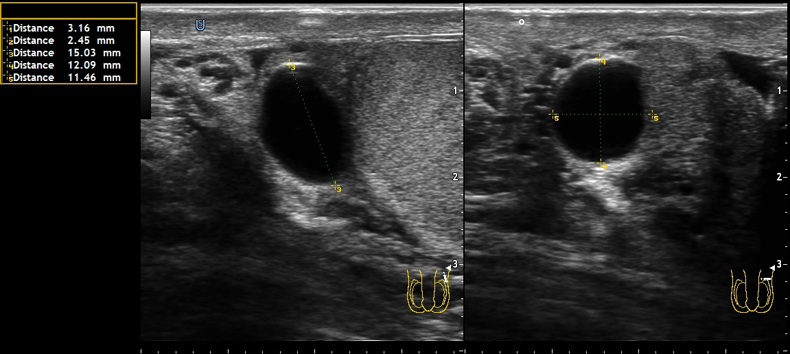
Cyst of the epididymis

General System Parameters

These parameters are consistent across the entire SONUS product line—regardless of the number of ports or sensor configuration.
PARAMETER VALUE
Frequency range (broadband system) 1–14 MHz
Number of levels on the gray scale at least 256
Dynamic Range at least 263 dB
Focusing the received signal At least 7 controllable zones, digital dynamic with apodization
Scanning depth at least 40 cm
Measurement range for linear dimensions 1–300 mm
Touchscreen diagonal at least 23.8″
Image Archive 500 GB or more of built-in storage + patient database
Enlarge the image up to 420× (optical up to 13.4×, digital up to 32×)
Frame rate over 400 frames per second
Size of the film loop 2048 frames (optional: 4096)

Display Modes

B-modes and image enhancement technologies
B, B/B, 4B –two-dimensional grayscale modes;
THI – B- and M-modes on a tissue harmonica;
Inv H – B- and M-modes on an inverse harmonica.

M-Modes
M (B/M) – time-scanned M-mode and combined B/M-mode;
MA – anatomical M-mode;
MC (MCD, MPD, MDF, MTD) – M-mode color mapping.

Color Doppler Mapping
CD (B/CD) – color mapping of blood flow velocity;
PD (B/PD) – energy mapping of the Doppler signal;
DF (B/DF) – color mapping of dynamic flow;
TD (B/TD) – tissue doppler color mapping;
VP – pulsed blood flow color mapping.

Spectral Doppler Modes 
D (PW) – pulsed-wave spectral doppler;
CW – continuous-wave doppler.

Combined and multi-screen modes
B/D – pulse-wave duplex;
B/CW – continuous-wave duplex;
CD(PD,DF,TD)/D(PW) – triplex mode;
CD(PD,DF,TD)/CW – alternating triplex mode;;
CD(PD,DF,TD)/CD(PD,DF,TD) – alternating dual-window modes;
4CD(PD,DF,TD) – four-window display modes;
CD(PD,DF,TD)/MCD(PD,DF,TD) – combined color mapping of B- and M-modes.

Elastography and Quantitative Modules
SE (B/SE) – shear-wave elastography mode;
CE (B/CE) – compression elastography mode;
AC –color mapping and quantitative assessment of the ultrasound attenuation coefficient in tissue;
E-AS – automatic assessment of vascular wall elasticity;
E-IMT – automatic assessment of intima-media thickness.

The SONUS ultrasound scanning system complies with the requirements of the Technical Regulation on Medical Devices for Class IIa devices in terms of the potential risk associated with its use

The SONUS ultrasound scanning system meets the safety requirements of DSTU EN 60601-2-37 and DSTU EN 60601-1 for Class I devices in terms of protection method and Type BF protection against electric shock.

The SONUS ultrasound scanning system complies with the EMC requirements of DSTU EN 60601-2-37 and DSTU EN 60601-1-2 for Class A, Group 1 devices according to DSTU EN 55011:2014.

The system is equipped with a function to display ultrasound power, mechanical, and thermal indices, which allow monitoring the level of ultrasound energy transmitted to the patient.

In some modes, automatic transmission power limitation is enabled.

The maximum surface temperature of the transducer applied to the patient’s body and operating in radiation mode complies with the requirements of DSTU EN 60601-2-37 and does not exceed 43 °C.

Modifications

SONUS One

1 sensor port

Basic examinations of the abdominal cavity, small organs, superficial vessels, and the musculoskeletal system

  • 6–15 MHz linear sensor included

Popular

SONUS Plus

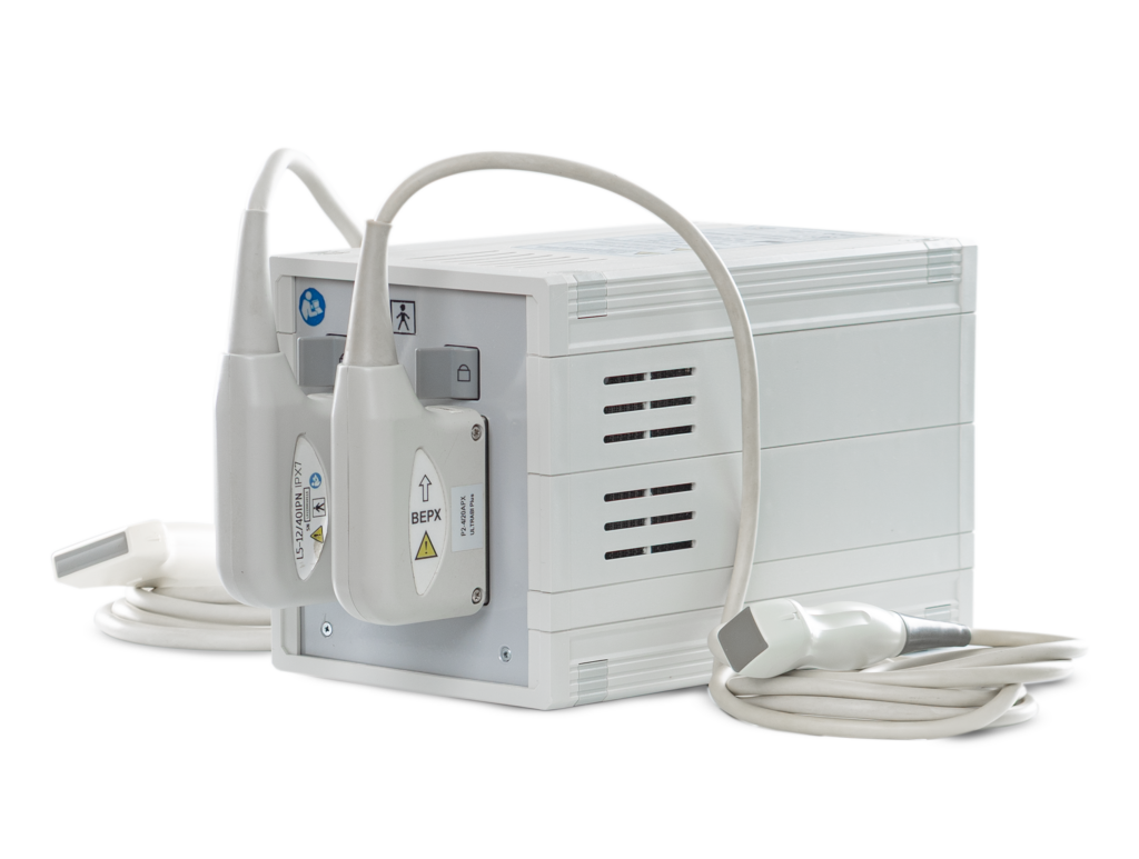
2 sensor ports

They enable advanced examinations of the abdominal organs, blood vessels, and in the fields of obstetrics and gynecology and urology

  • ЛLinear (6–15 MHz) + convex (2–5 MHz) sensors included
  • Simultaneous connection of two sensors

SONUS Pro

2 sensor ports

Recommended for a multidisciplinary clinic offering a full range of tests

  • Linear (6–15 MHz) + convex (2–5 MHz) transducers included
  • Also supports shear-wave elastography for quantitative assessment of tissue stiffness
ParameterSONUS OneSONUS PlusSONUS Pro
Number of sensor ports122
Touchscreen, no less23.8″23.8″23.8″
6–15 MHz linear sensor included✓✓✓
2–5 MHz convex transducer included—✓✓
Connecting a 2–4 MHz phased sensor✓✓✓
Color/Energy Doppler✓✓✓
Compression elastography✓✓✓
Shear-wave elastography (optional)——✓
System/Sensor Warranty24 / 12 month24 / 12 month24 / 12 мmonth
Cartavailable for order

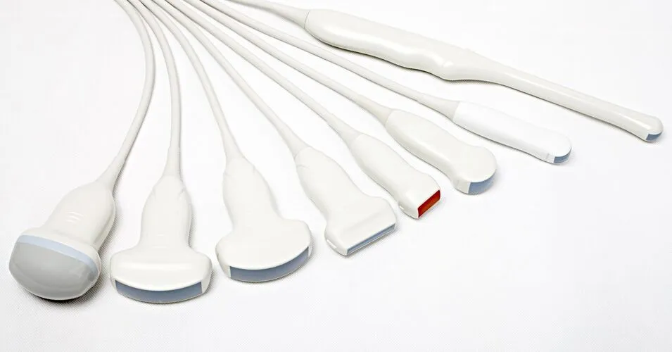
Sensors

50R; 1,5-5,0 MHz; 128 el)

(60R; 1,5-5,0 MHz; 128 el)

(50R; 3,0-7,0 MHz; 192 el)

(50R; 3,0-7,0 MHz; 192 el)

(38 mm; 4,5-12,0 MHz; 192  el)

(38 mm; 4,5-10,0 MHz; 128 el)

(38 mm; 5,5-13,0 MHz; 192 el)

(55 mm; 5,0-12,0 MHz; 192 el)

(900; 1,7-4,0 MHz; 64 el)

(11,5R; 3,6-9,0 MHz; 128 el) 

(11,5R; 3,6-9,0 MHz; 128 el)

(11,5R; 3,6-9,0 MHz; 192 el)

Request a call

Get a consultation

Request a call

Get a consultation