A reliable device from the manufacturer with support throughout its entire period of use
Free delivery and installation
24 months official warranty
Premium quality for professional use
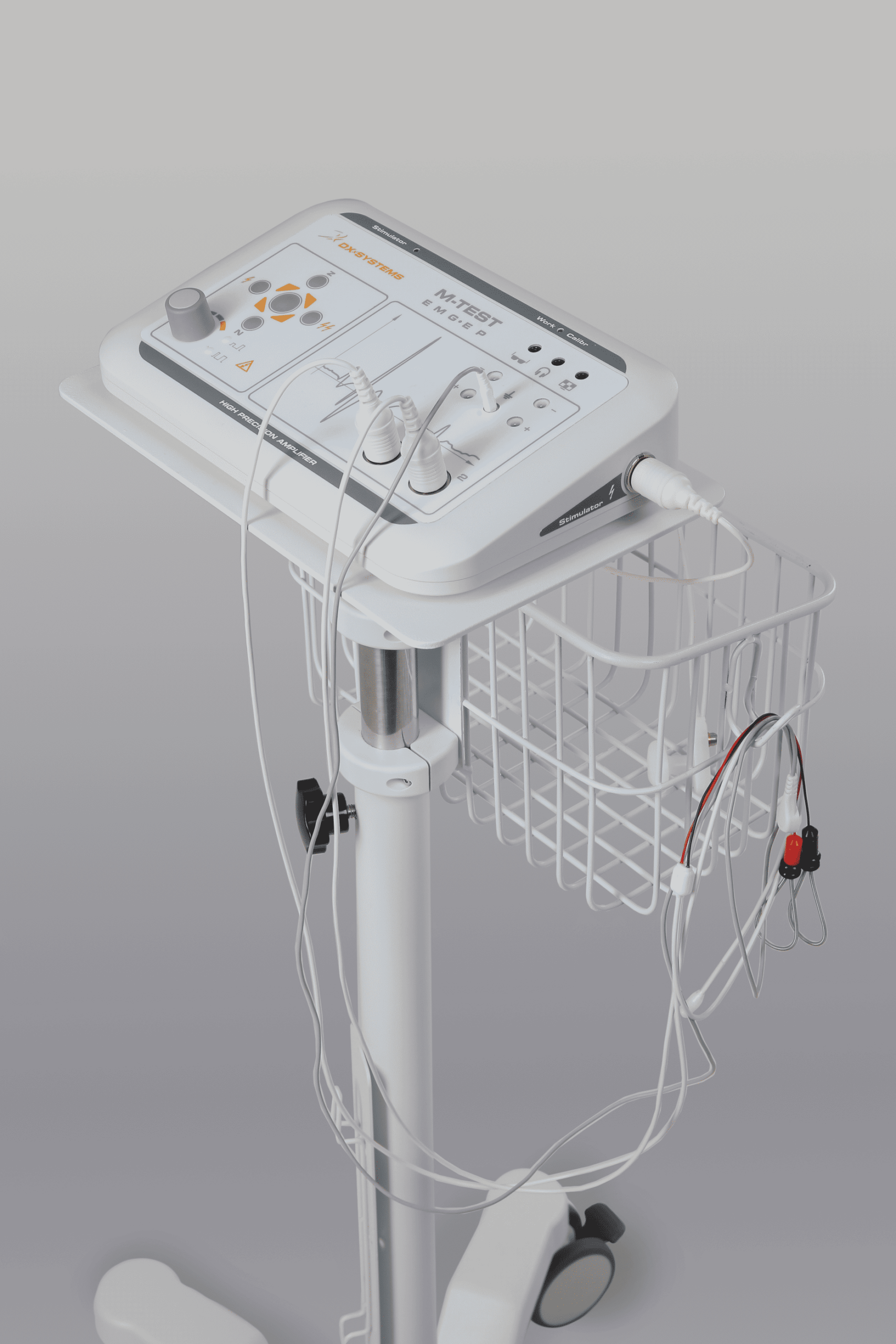
Comprehensive Solution for Neuro-Muscular Diagnostics A versatile system for evaluating the neuromuscular system through the recording of muscle and peripheral nerve electrical potentials.
Designed for use in neurology, surgery, rehabilitation, sports medicine, and clinical research.
Capability to monitor multiple muscles and nerves simultaneously
Comparative assessment of muscle function
Assessment of auditory/visual/cognitive function
Models & Configurations:
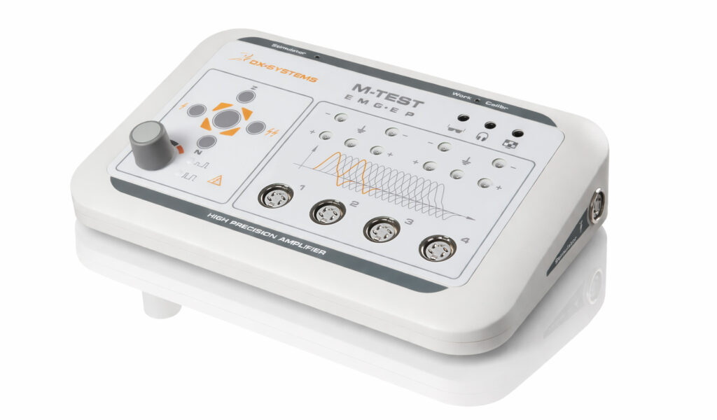
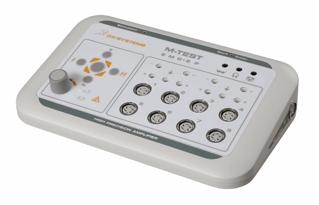
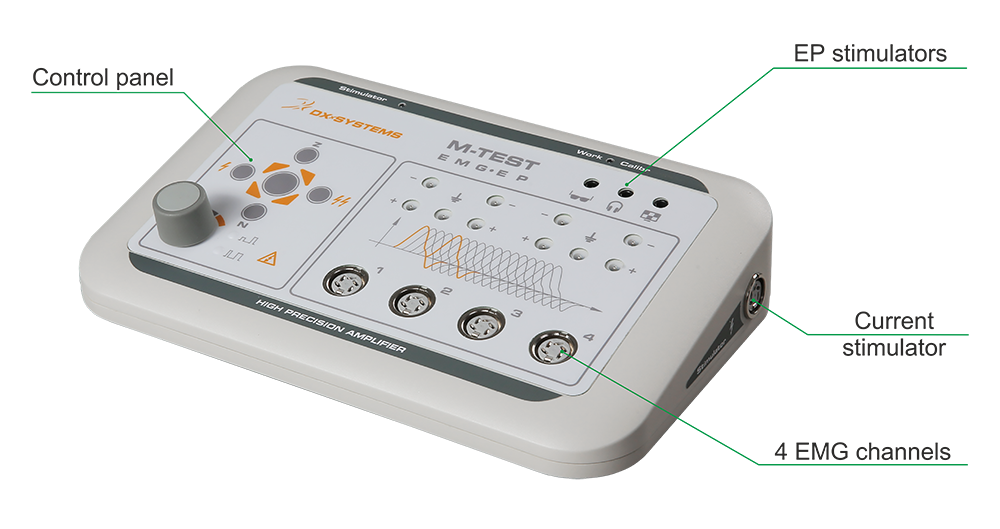
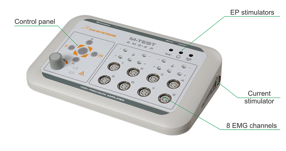
2 / 4 / 8 acquisition channels
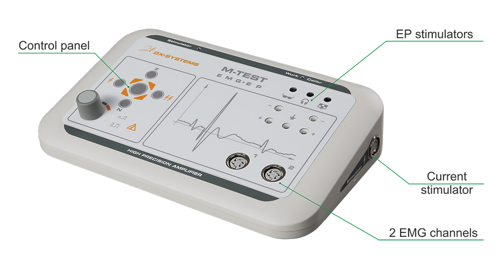
Integrated current stimulator
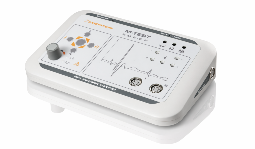
Intuitive controls with LED indicators
Portable and ergonomic design
Power Bank support for challenging environments
Functionality:
Broad range of EMG and NCS methodologies
Structured patient database with easy access to studies and reports
Direct access to key functions via device panel, footswitch or keyboard
Predefined test templates for routine workflows
Real-time signal filtering with adjustable parameters
Stimulation artifact suppression
Automated report generation based on clinical norms
Flexible reporting options
Automatic and manual software update checks
Safety & Compliance:
CE certified, conforming to European safety standards and the Medical Device Technical Regulation
Designed for reliable, efficient, and user-friendly operation.
🟠 Surface EMG – general assessment of muscle electrical activity.
🟠Stimulation EMG (Nerve Conduction Studies) – evaluation of conduction velocity and reflex responses.
Methods:
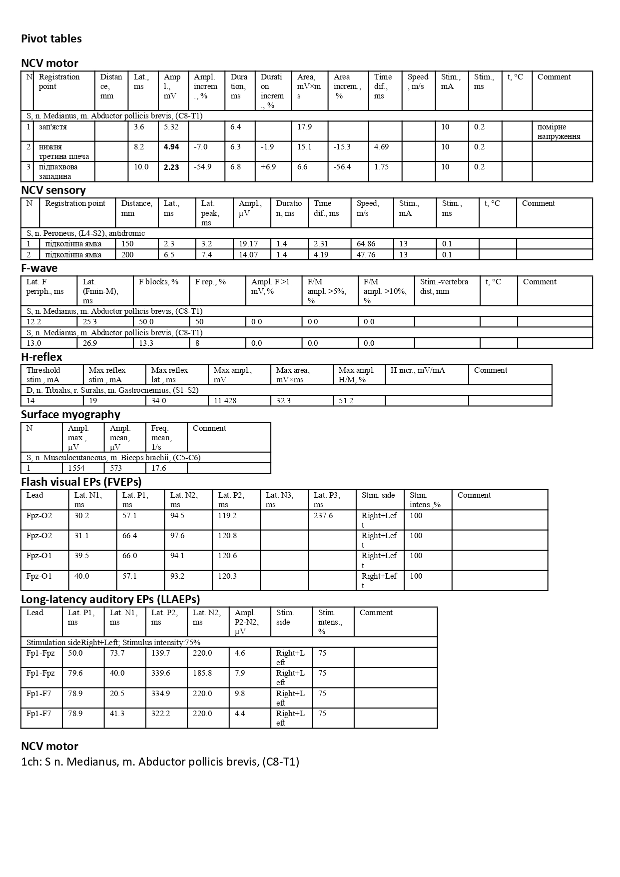
Motor / Sensory NCS
F-wave
H-reflex
Blink reflex
M-wave decrement test
Repetitive nerve stimulation
Inching test
Magnetic stimulation
🟠Needle EMG – precise local diagnostics of muscle activity.
Methods:
Spontaneous activity
Motor unit potentials
Interference pattern analysis
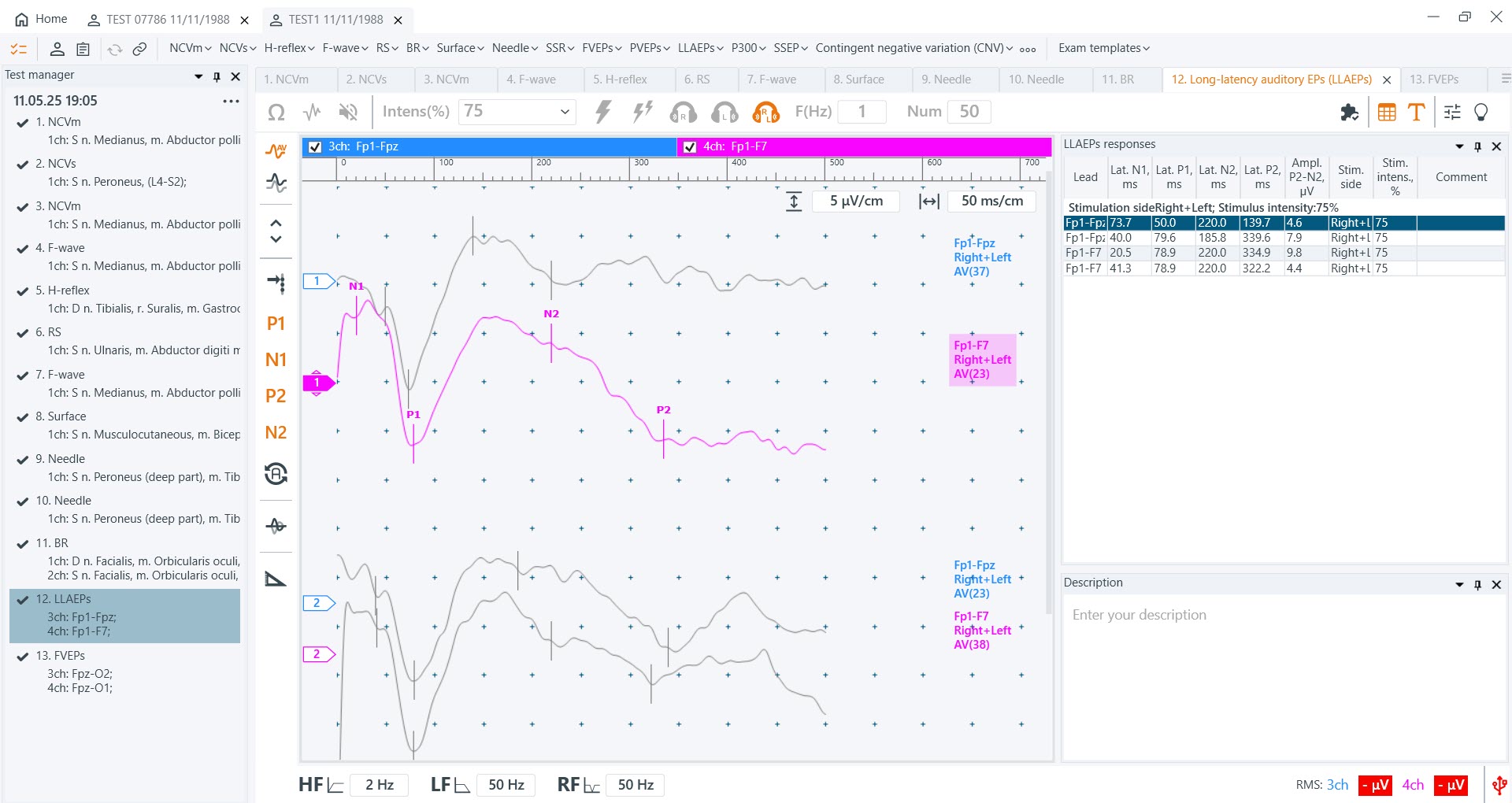
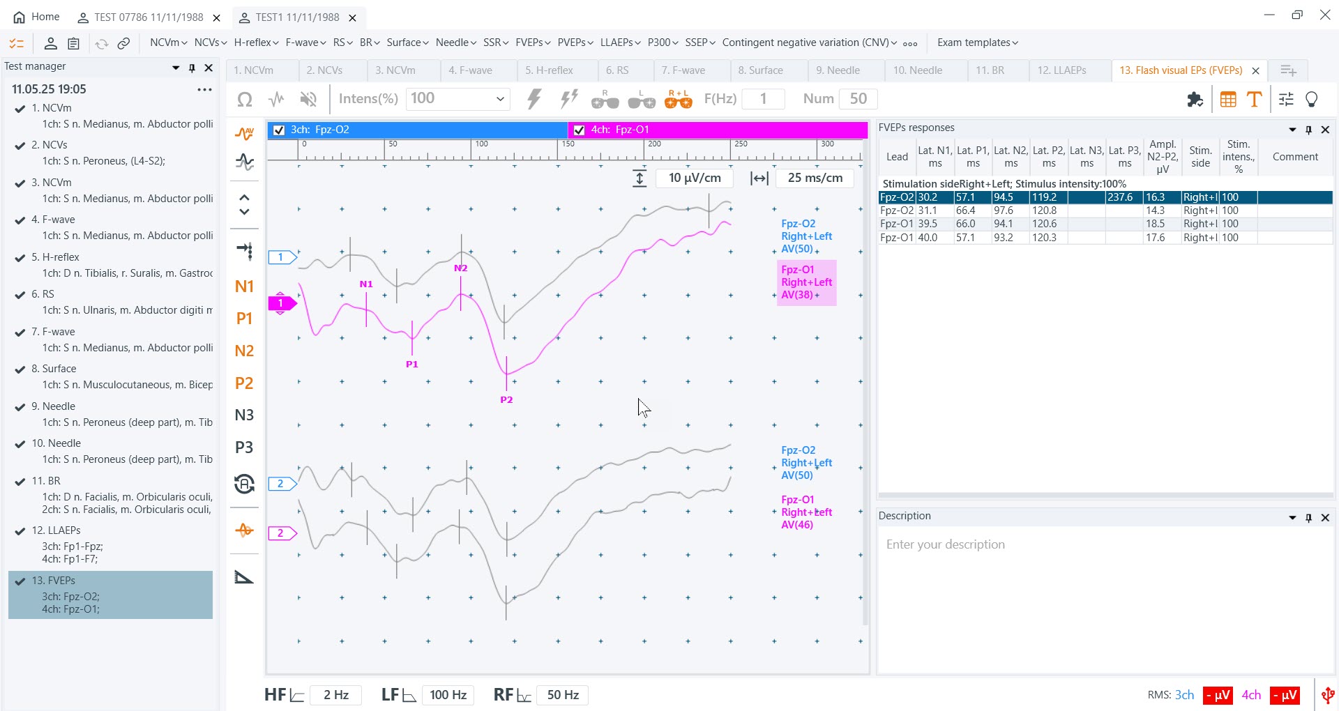
🟠Evoked Potentials (EPs) – evaluation of sensory, cognitive, and autonomic functions
Methods:
Somatosensory EPs (SSEPs)
Visual EPs (flash, pattern)
Auditory EPs (BAEP, long-latency)
Cognitive EPs: P300, CNV
Autonomic skin response (SSR)
Base set:
Hardware unit
Software suite
Set of electrodes and accessories
Technical documentation
Optional Set:
Evoked Potentials – enables advanced EP testing modes
Convenience & Mobility Solutions:
Mobile cart / desktop mount / portable workstation table
Carrying case for device, laptop, and accessories
Model
EMG
channels
Equipment set
M-TEST ONE-8
8
Base
M-TEST ONE-8 EP
8
Base + evoked potentials: short-latency somatosensory, long-latency auditory and visual, cognitive
M-TEST ONE-4 EP
4
Base
M-TEST ONE-4 EP
4
Base + evoked potentials: short-latency somatosensory, long-latency auditory and visual, cognitive
M-TEST ONE-2
2
Base
M-TEST ONE-2 EP
2
Base + evoked potentials: short-latency somatosensory, long-latency auditory and visual, cognitive
Parameter
Value
Number of EMG channels
2 / 4 / 8
Input signal range
10 – 60,000 µV
Input impedance
≥ 100 MΩ
Voltage of internal noise of complexes, brought to the input – EMG channels – VP channels in the frequency range up to 100 Hz – VP channels in the frequency range up to 3 kHz
≤ 5 µV ≤ 1 µV ≤ 5 µV
Sampling rate
16000 Hz
Frequency range
0.2 Hz – 3.0 kHz
Low-frequency filter time constant
≥ 1.0 s
Attenuation coefficient of a common-mode signal at a frequency of 50 Hz
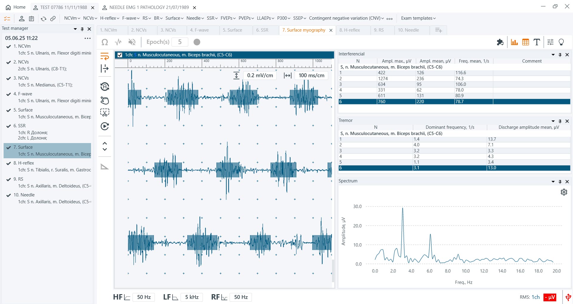
Main parameters of interference EMG: frequency of total muscle electrical activity, maximum signal amplitude, average signal amplitude. For orthopedic dentistry, an additional analysis of the chewing test is provided. In pain studies, the investigation of voluntary muscle activity is used.
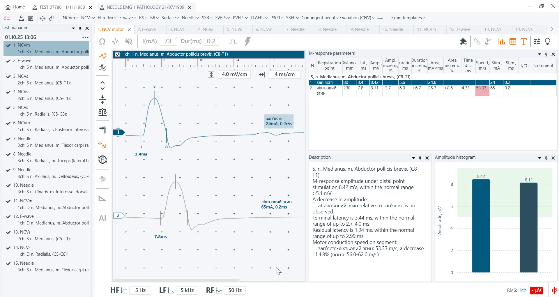
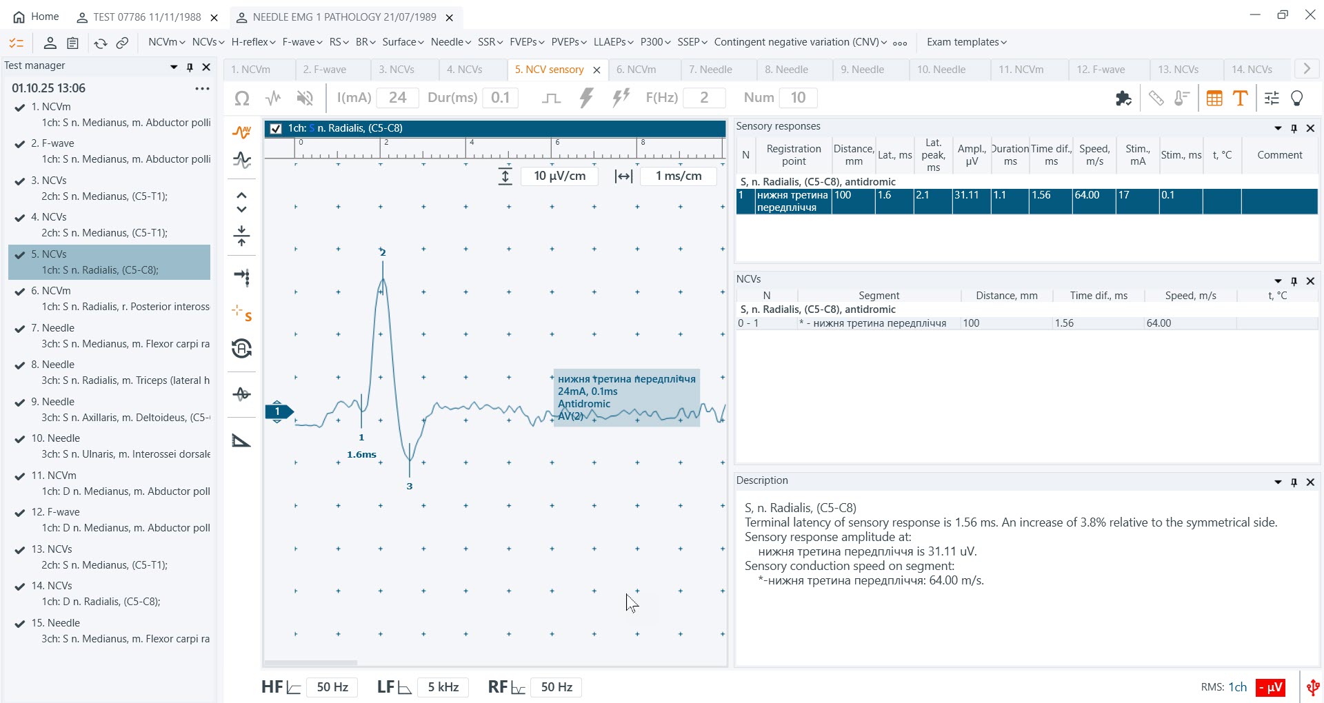
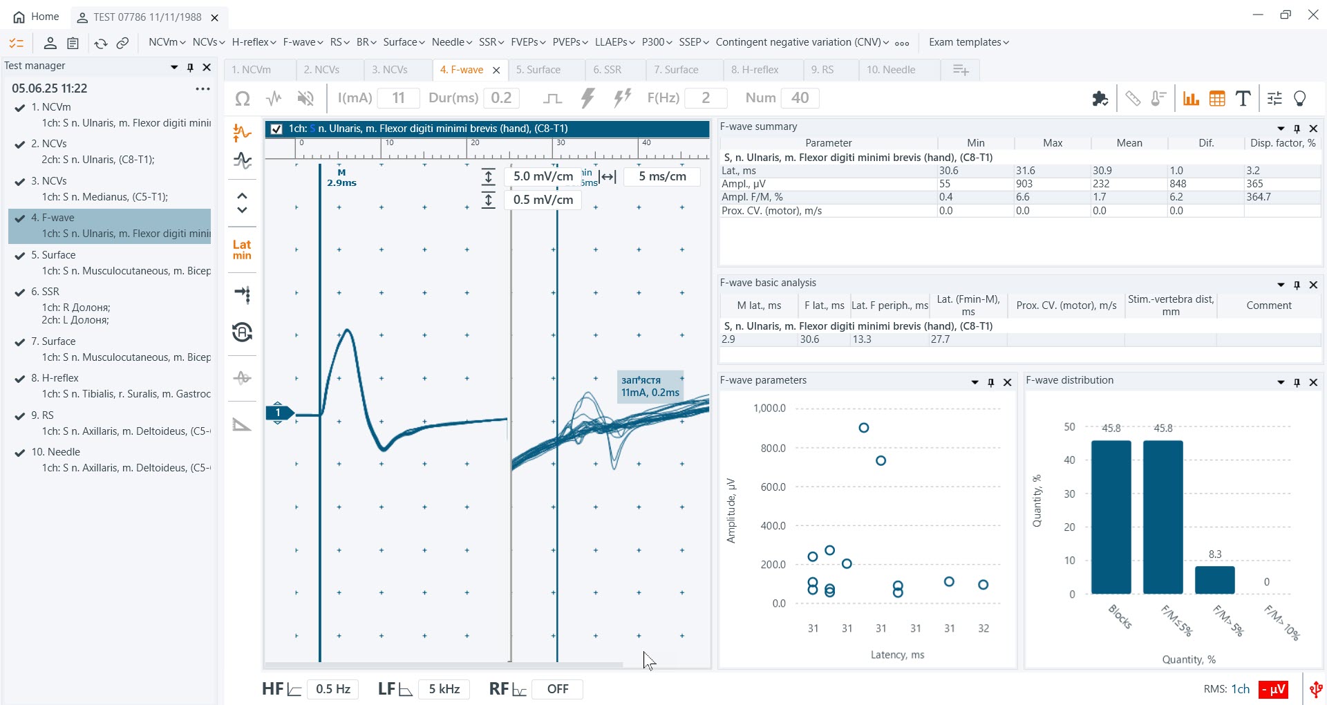
Stimulation EMG
The use of current stimulation ensures high accuracy in the study of nerve conduction. Muscle response or electrical activity is measured using a current stimulus of precise duration and amplitude. Early responses (M-response) or late responses (F-wave, blink reflex, H-reflex) allow determining the level and localization of peripheral neuromotor system lesions. Sensory and motor fibers can be assessed separately by measuring sensory and motor conduction velocity. Therefore, it is fair to note that this group includes basic methods applied in the differential analysis of neuromuscular diseases.
A special type of response analysis to stimulation is analysis using rhythmic stimulation. The decrement test is performed to evaluate the reliability of neuromuscular transmission. The tetanic contraction test and pharmacological loading can be used to detect myasthenia and Lambert-Eaton myasthenic syndrome.
Needle EMG
Needle electromyography (local EMG) studies the functional state of muscles at rest and under voluntary load. The results of needle EMG may be useful in primary muscle diseases, which differ from those observed in nervous system disorders. Based on the results of a comprehensive ENMG study, the level of peripheral neuromotor system damage, the nature, degree, and extent of the pathological process are determined, and in repeated studies – the effectiveness of therapy.
Methods of recording evoked potentials differ significantly from other studies conducted within EMG.
They address specific tasks. Visual evoked potentials can be used to confirm damage to the visual tract, while auditory evoked potentials are used to assess hearing and the function of nerve pathways from the ear to the brain, helping to detect even hidden disorders at early stages. These types of EPs can be sensitive to dysfunctions that cannot be detected solely through physical examinations or MRI.
Somatosensory evoked potentials are often used in neuromonitoring to assess spinal cord function during surgery.
Galvanic skin response is an indicator of autonomic nervous system activity.
The M-TEST ONE software allows convenient work with databases – in addition to all patient information, recorded electromyograms, and their processed results, it provides a full set of functions for comfortable work with electronic patient files and large-volume information archives.
To optimize the preparation of medical documentation, report generation is highly automated. Examination results can be printed, saved in digital format, or transmitted via email or local network.
Mathematical and statistical processing of electromyograms is automatically presented in a format suitable for forming a conclusion.
The software provides functions for exporting and importing all electromyographic examination data with the possibility of further information exchange with other specialists for practical use or scientific research.