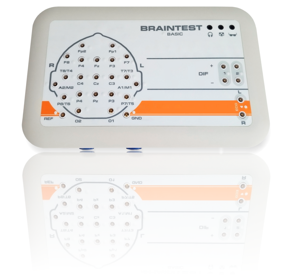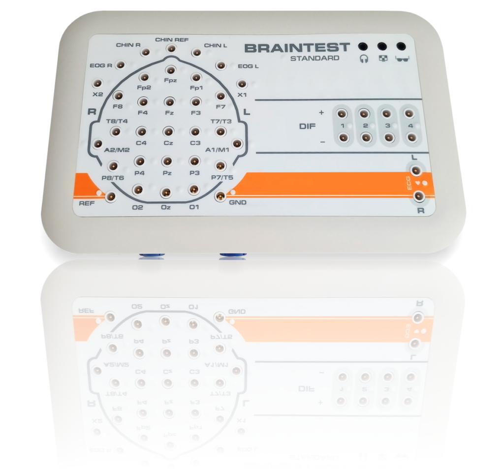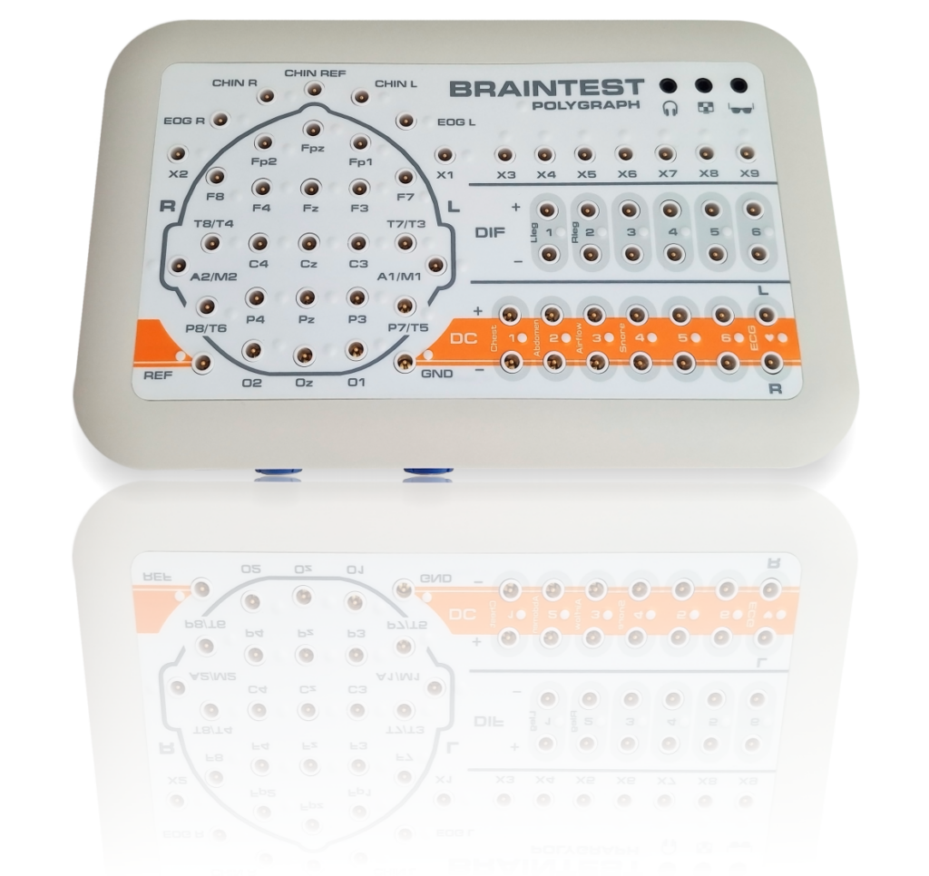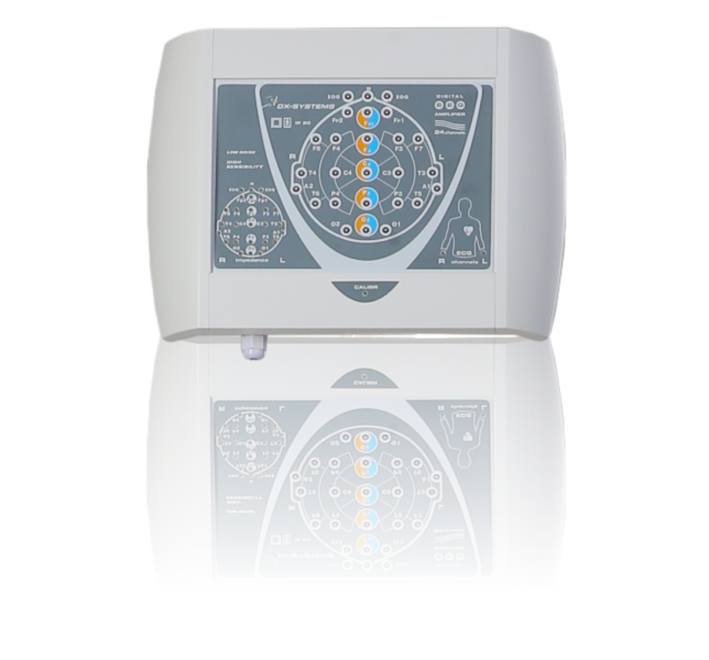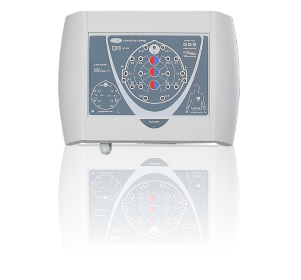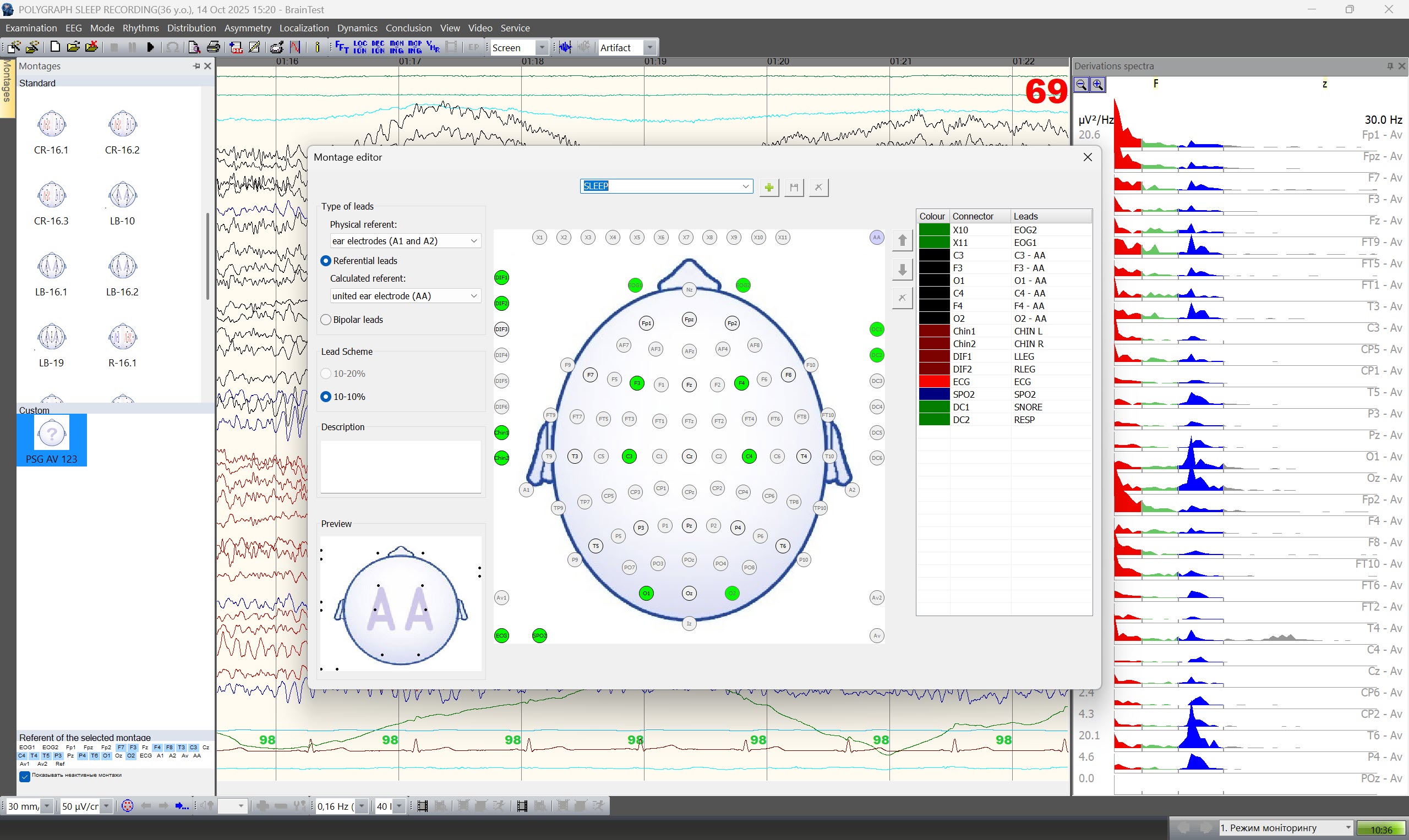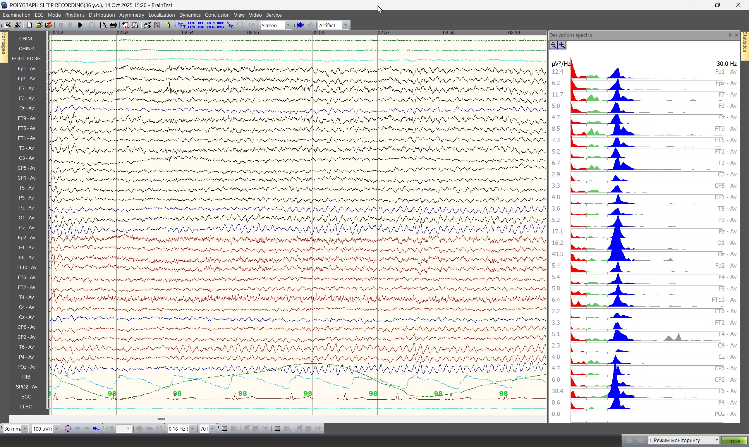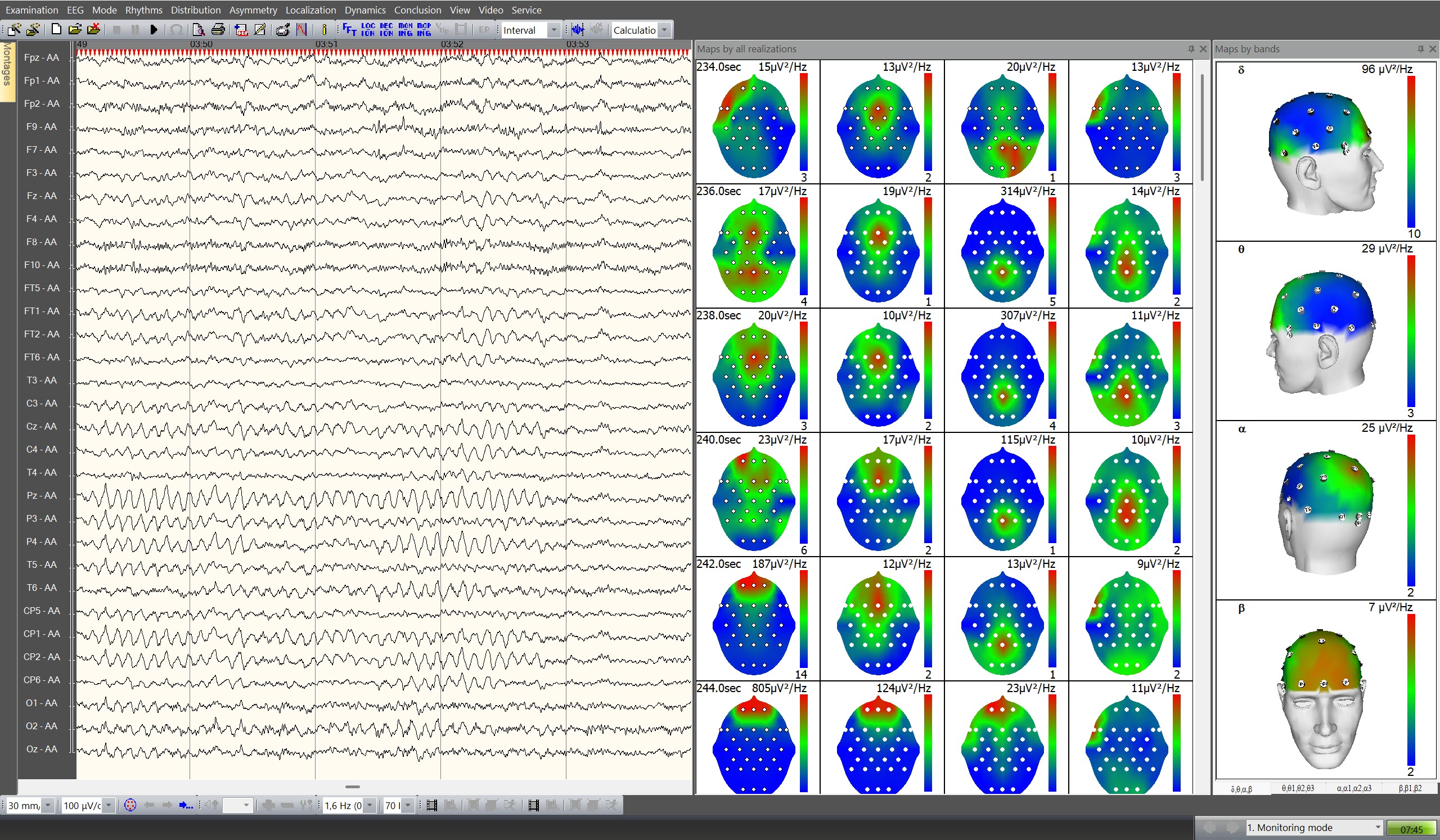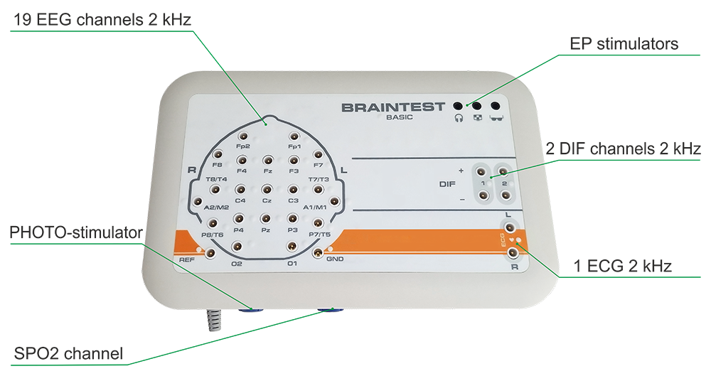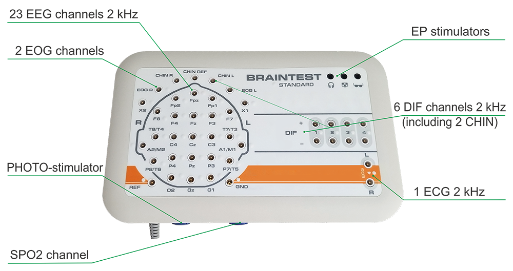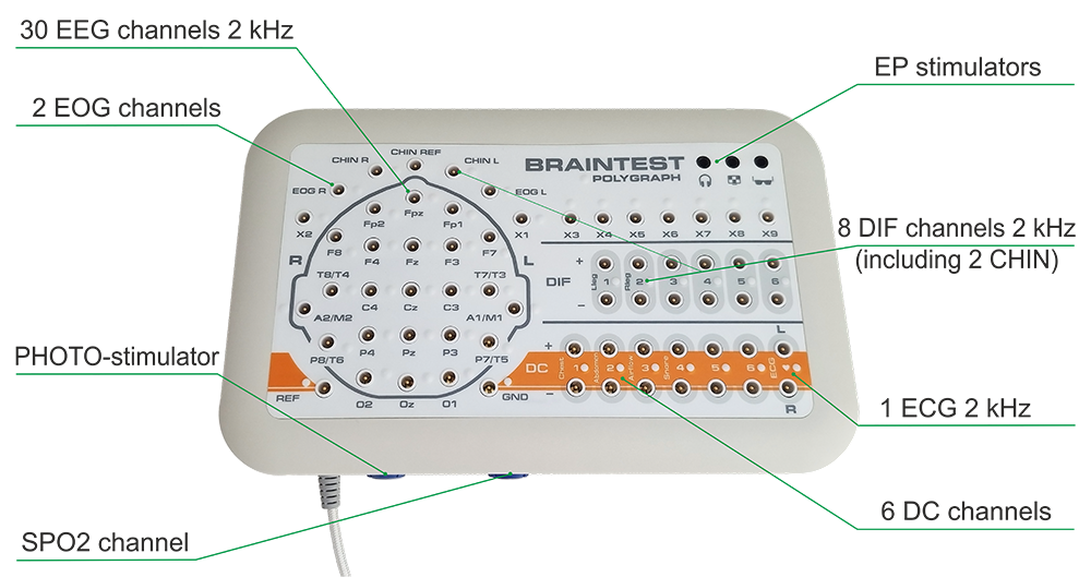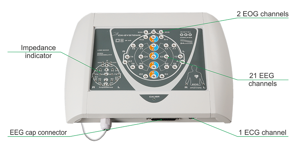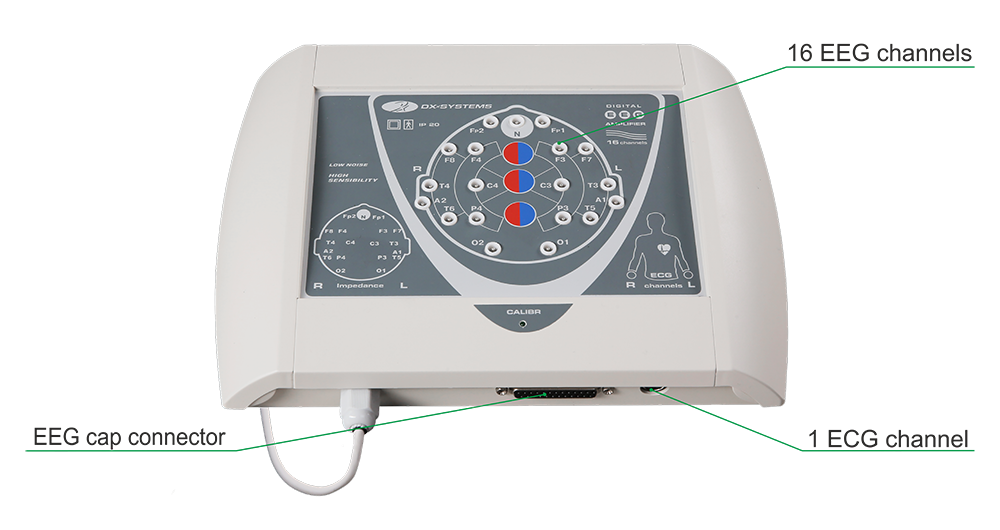

 EEG system BRAINTEST series – base equipment set EEG system BRAINTEST PLUS series – base equipment set
Models:
Additional modules|
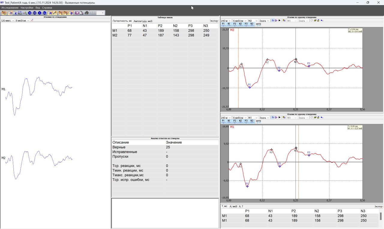
EVOKED POTENTIALS |
Long-latency auditory and visual, cognitive |
|
VIDEO MONITORING |
Long-term synchronous EEG and video recording (up to 72 hours) |
Additional modules, at the customer’s request, are supplied with all EEG models of the BRAINTEST and BRAINTEST PLUS series.