 Skip to content
 A reliable device from the manufacturer with support throughout its entire period of use


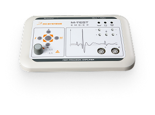
Non-invasive study of central hemodynamics of blood flow in the brain, limbs, and internal organs.
Used in neurology, scientific research, and diagnostic practice.

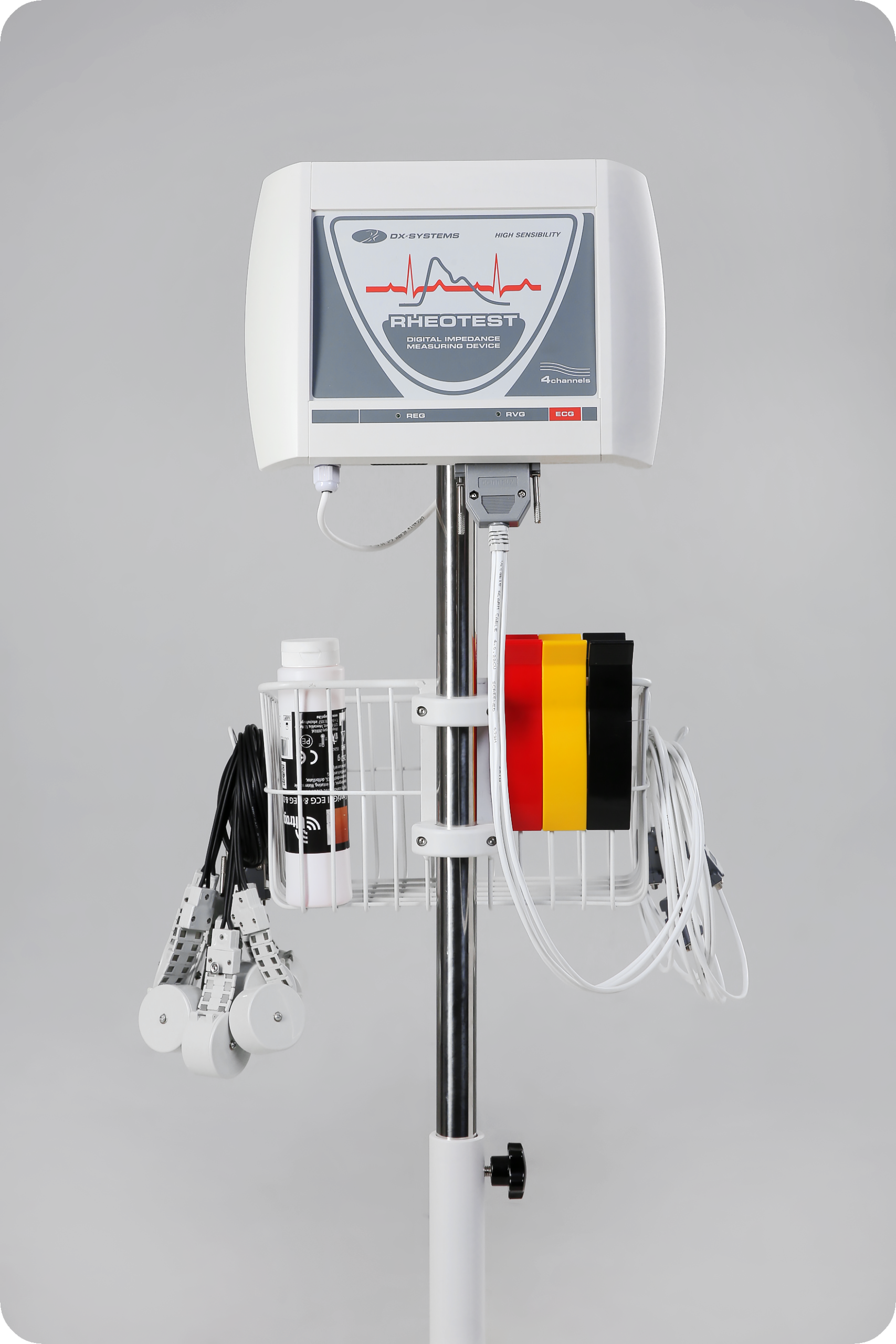
Complies with Technical Regulation on Medical Devices No. 753. Designed for professional, convenient, and efficient work.
🟠 Rheoencephalography (REG) – study of cerebral blood flow
🟠 Rheovasography (RVG) – analysis of limb circulation
🟠 Rheocardiography (RCG) – tetrapolar chest rheography using Kubíček’s method, integral rheography using Tyshchenko’s method
🟠 Rheohepatography (RHG) – study of liver blood flow
🟠 Rheopulmonography (RPG) – analysis of pulmonary circulation
🟠 Rheorenography (RRG) – assessment of renal blood flow system
🟠 Option 1: RHEOTEST REG
🟠 Option 2: RHEOTEST REG+RVG
🟠 Option 3: RHEOTEST MAX (REG+RVG+RKG+RGG+RPG+RRG)
| Parameter | Value |
|---|---|
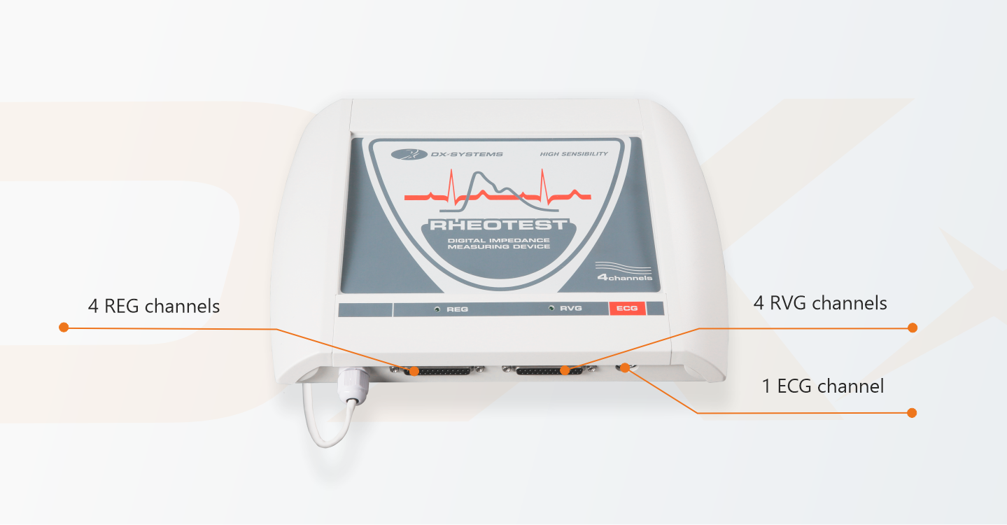
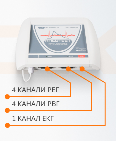
| Number of rheographic channels | 4 |
| Number of ECG channels | 1 |
| Base impedance measurement range | 10 – 500 Ω |
| Dynamic impedance measurement range | 0.01 – 0.5 Ω |
| Noise level reduced to the input | ≤ 0.003 Ω |
| Time constant | ≥ 0.4 s |
| Probing frequency | 4 / 28 / 56 / 112 kHz |

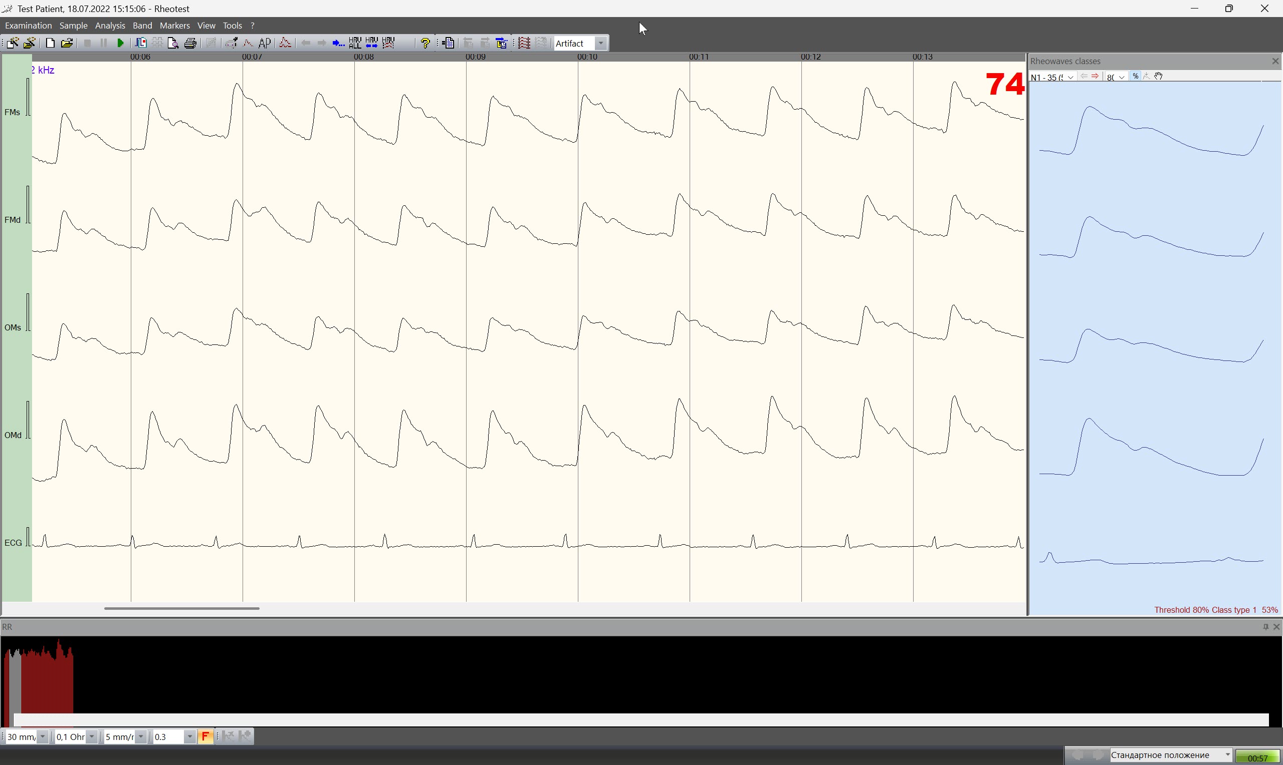
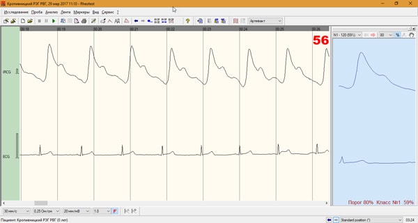
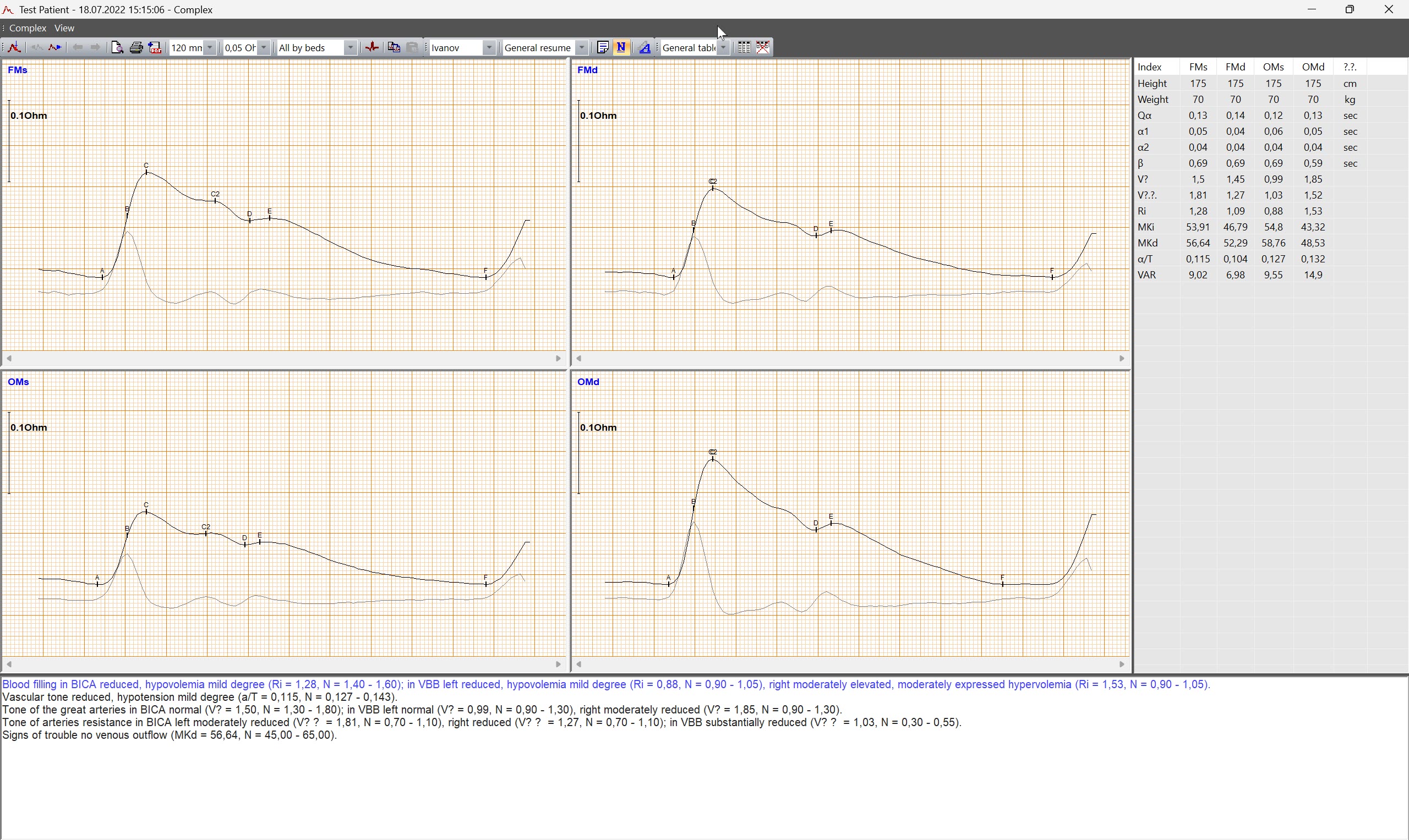
The capabilities of the RHEOTEST rheograph allow the physician to obtain maximum diagnostically significant information: during the registration of the rheogram, automatic detection and classification of rheographic complexes is performed, along with generation of the differential rheogram and determination of the current heart rate value.
The ease of visual assessment of rheographic wave parameters is achieved by displaying the curve on a millimeter grid.
For convenience in conducting examinations, a protocol function is provided, which can include any number of functional tests.
Even during signal registration, primary analysis of one of the recorded complexes can be performed by selecting any complex for comparison with another—for example, one included in a different functional test, or a similar test from an examination conducted a month earlier.
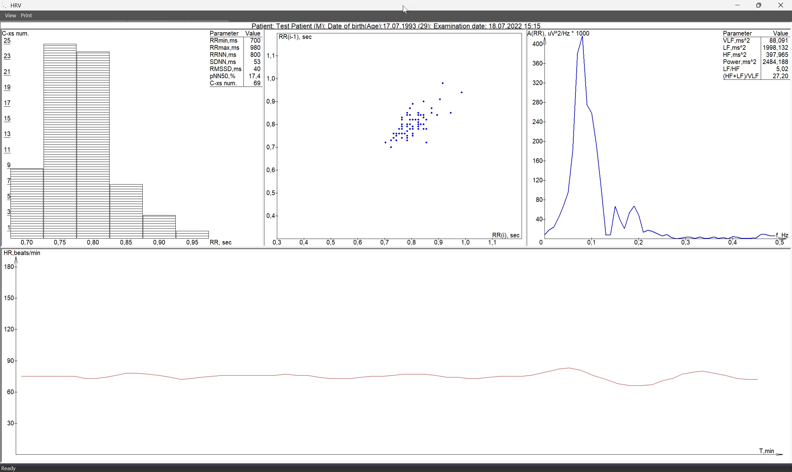
The rhythmogram window allows instant identification of positions on the rheographic recording that require special attention. By clicking on a section of the rhythmogram, precise positioning on the rheographic curve is performed at the moment corresponding to a specific heart rate. On the rhythmogram, the following are color-marked: current state; segments unsuitable for analysis and marked as artifacts.
While viewing the rheogram, the display of leads can be customized according to the user’s preferences, for example, changing their order or disabling them.
During rheogram analysis, you can select a set of characteristics depending on the vascular pool being studied.
The rheograph software automatically detects and classifies rheocomplexes using markers (adjustable manually) and calculates amplitude-time parameters.
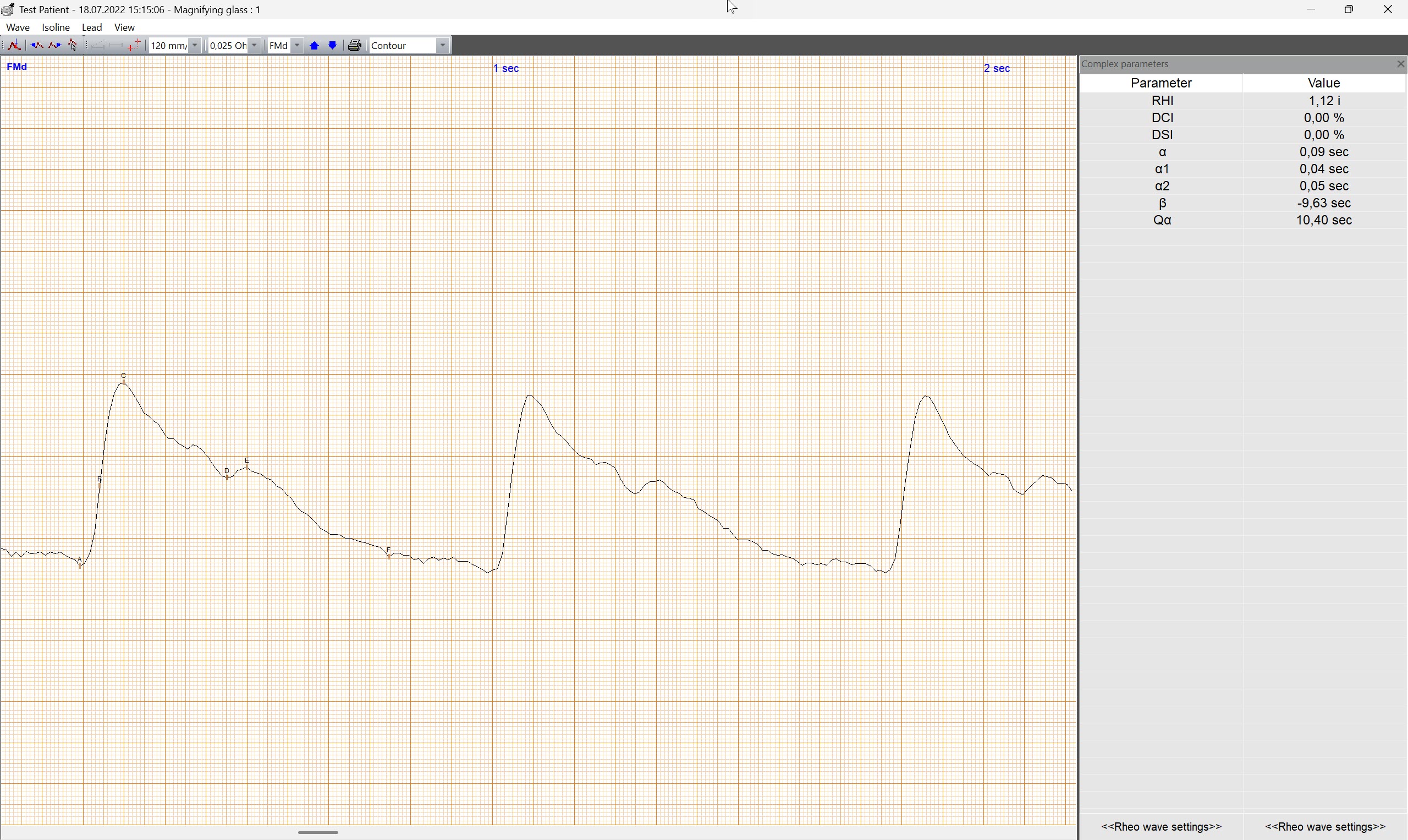
The “Magnifier” window allows analysis of the selected rheographic complex. Simultaneously with rheogram registration, rheographic complexes are automatically highlighted, the differential rheogram is displayed, and the current heart rate is determined.
Any of the recorded complexes can be analyzed and selected into a fragment group for comparison with any other part of the recording. The set of characteristics is chosen depending on the vascular pool under study. The “Complex” window displays the rheocomplex across all channels and a summary table of amplitude-time parameters. You can choose how to display the rheogram: separately by each channel, in pairs from the same vascular pool, or from the same side.
The program evaluates the nature and dynamics of rheogram changes both during a standard study and while recording functional tests.
For the analysis of the entire recorded rheogram, the ribbon can be scrolled either automatically or manually. An interesting fragment can be located and viewed using the rhythmogram. During rheogram viewing, it is possible to change the order of lead display, swap them, or disable any of them.
Heart rate variability analysis is provided: time-domain (statistical), wave structure analysis of heart rate, scattergram, and variational pulsometry according to Baevsky. Automatic description of the rheogram is generated according to the selected method and corresponding age norms. At the same time, the system allows the user to create their own system of norms and decision-making rules for any methodology.
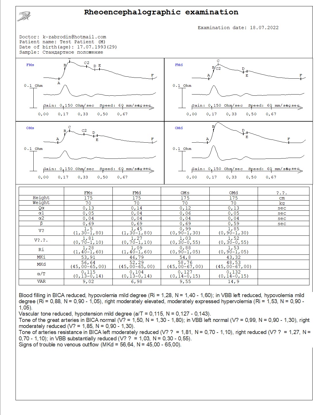
The physician decides on the content and format of the printed report. This may include a rheocomplex with amplitude-time characteristics and an automatic description generated according to the selected method and considering age norms.