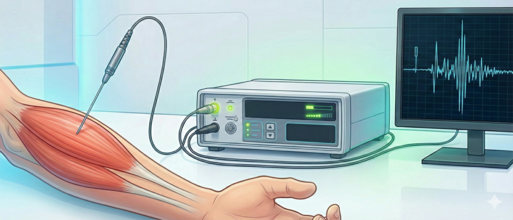
FEATURES OF AMPLIFIERS FOR NEEDLE ELECTROMYOGRAPHY RECORDING
Zabrodin K. Yu., Geletka O. O.,
postgraduate student, candidate of medical sciences, medicine category
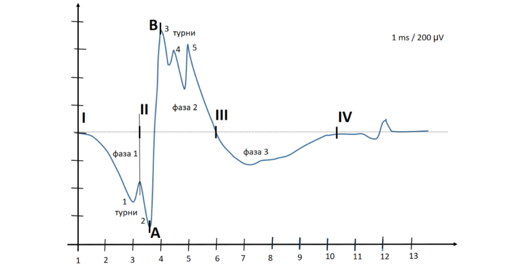
MAIN CHARACTERISTICS OF MOTOR UNIT POTENTIALS RECORDED WITH A CONCENTRIC NEEDLE ELECTRODE
Zabrodin K. Yu., Geletka O. O.,
postgraduate student,
Candidate of Medical Sciences, Doctor of the Highest Category
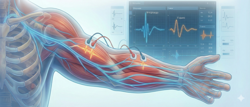
METHODS OF STIMULATION ELECTRONEUROMYOGRAPHY
Abstract
This document discusses the main purpose of stimulation electroneuromyography. The importance of conducting such an examination for a wide range of pathologies is emphasized. The main criteria for prescribing this examination to patients of different age groups are given. The main methods of stimulation EMG, as well as the parameters of the device settings during the examination, are considered.
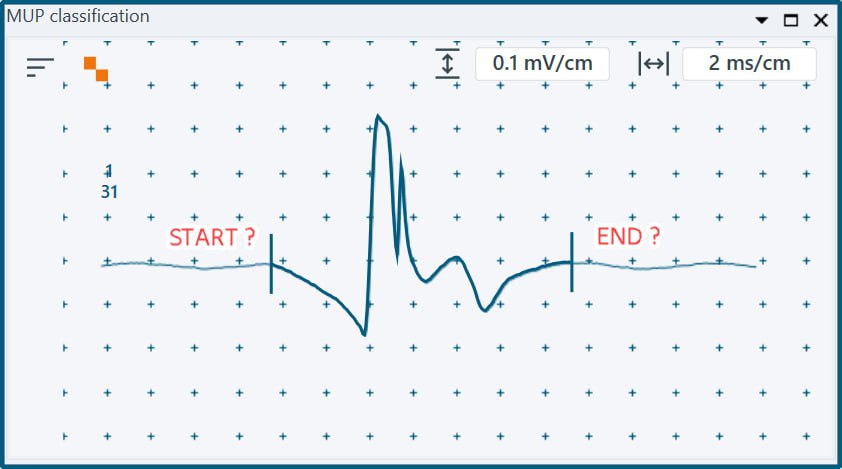
METHODS FOR AUTOMATIC LABELING OF MOTOR UNIT POTENTIALS RECORDEDWITH THE USE OF A CONCENTRIC NEEDLE ELECTRODE
Abstract
The article reviews the main methods for determining motor action unit potential (MUAP) duration in electromyography. Various algorithms, such as the Turku 1 method, the Turku 2 method, the
Stolberg method, and the Nandedkar method, which are used to determine the MUP limits are analyzed. The advantages and disadvantages of each approach are described, as well as their practical applicability in clinical and scientific electromyography.